"All we have discovered is that it starts with a single individual - always a child - and then spreads explosively, like the formation of crystals round the first nucleus in a saturated solution." Arthur C. Clarke (Childhood's End, 1953)
Facility
 Our
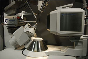
X-ray facility owns two single crystal X-ray diffractometers: (i) one Xcalibur 3 diffractometer from Oxford
Diffraction equipped with a 4-circle Kappa goniometer (KM4) and
with a Sapphire 3 CCD detector. (ii) one state-of-the-art SuperNova diffractometer from Rigaku Oxford Diffraction (formerly Agilent Technologies) which combines two hi-flux micro-focus sources (Mo and Cu) equipped with a large area CCD Atlas detector. Both instruments are equipped with
a Cryojet low-T device from Oxford
Instruments so that data can be collected at low temperature (110
K), which offers several significant benefits (reduction of thermal
motion, higher data resolution, data collection for relatively air/moisture/temperature
sensitive crystals). Temperature can also be tuned between 100 and 300
K for special cases (e.g., destructive and non-destructive phase transitions).
Data collection/reduction is processed with the intuitive CrystAlisPRO
software on one PC workstation (Windows). Softwares are generally updated
on a six month basis. The X-ray lab has two extra PC workstations used
for CSD
(Cambridge Structural Database) searches and structure solution &
refinement.
Our
X-ray facility owns two single crystal X-ray diffractometers: (i) one Xcalibur 3 diffractometer from Oxford
Diffraction equipped with a 4-circle Kappa goniometer (KM4) and
with a Sapphire 3 CCD detector. (ii) one state-of-the-art SuperNova diffractometer from Rigaku Oxford Diffraction (formerly Agilent Technologies) which combines two hi-flux micro-focus sources (Mo and Cu) equipped with a large area CCD Atlas detector. Both instruments are equipped with
a Cryojet low-T device from Oxford
Instruments so that data can be collected at low temperature (110
K), which offers several significant benefits (reduction of thermal
motion, higher data resolution, data collection for relatively air/moisture/temperature
sensitive crystals). Temperature can also be tuned between 100 and 300
K for special cases (e.g., destructive and non-destructive phase transitions).
Data collection/reduction is processed with the intuitive CrystAlisPRO
software on one PC workstation (Windows). Softwares are generally updated
on a six month basis. The X-ray lab has two extra PC workstations used
for CSD
(Cambridge Structural Database) searches and structure solution &
refinement.
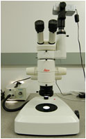 Crystals
and crystal quality are always inspected first under a stereomicroscope
(a Leica MZ75).
Careful examinations are always made for crystal morphology, crystal
color, crystal size, sample homogeneity, signs of twinning (under cross
polarized light) in order to maximize chances to find the most suitable
(single) crystal for X-ray structure determination. Equidimensional
crystals (e.g., blocks) are always best (to minimize absorption), and
large anisotropy in crystal shape (e.g., needles, plates) is less preferred.
Microsurgeries (using a razor blade) on lath- or rod-like crystals are
generally performed to minimize the anisotropy in shape. Microsurgeries
are also made when obvious twin domains can be separated. Crystals with
0.1-0.5 mm for each dimension are generally fine (crystals with 0.3-0.5
mm for each dimension are most often required for organic compounds).
Crystals
and crystal quality are always inspected first under a stereomicroscope
(a Leica MZ75).
Careful examinations are always made for crystal morphology, crystal
color, crystal size, sample homogeneity, signs of twinning (under cross
polarized light) in order to maximize chances to find the most suitable
(single) crystal for X-ray structure determination. Equidimensional
crystals (e.g., blocks) are always best (to minimize absorption), and
large anisotropy in crystal shape (e.g., needles, plates) is less preferred.
Microsurgeries (using a razor blade) on lath- or rod-like crystals are
generally performed to minimize the anisotropy in shape. Microsurgeries
are also made when obvious twin domains can be separated. Crystals with
0.1-0.5 mm for each dimension are generally fine (crystals with 0.3-0.5
mm for each dimension are most often required for organic compounds).
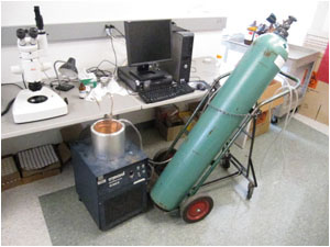
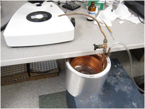
For crystals that are relatively air/moisture/temperature sensitive,
we use a simple homemade apparatus (see the pics below) generating a
cold N2 stream. The dewar, which is filled with liquid N2,
contains a copper coil aiming at cooling the input gas (room-temperature
N2 stream). The output gas (cold N2 stream) is
then directed onto the mounting stage of the microscope in order to
prevent crystal decomposition.
 |
 |
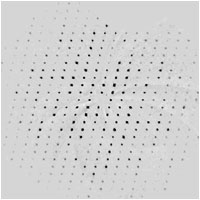
Crystals, which are thought to be suitable for X-ray structure determination, are mounted on the diffractometer under a N2 cold stream (typically 110 K). Picking the right crystal remains probably the most critical step in the process of X-ray structure determination (in other words, bad crystal quality nearly always lead to poor data quality and/or troubles in structural features). The diffraction pattern is then examined, and if satisfactory [e.g., crystals diffract at (sinΘ)/λ ≥ 0.59 Å-1, reflections appear sharp and unsplit], 10-15 frames (ω scans) are collected in order to determine the cell dimensions. At that stage, a quick search on the CSD can be beneficial for finding whether the crystal structure is known or not. For cases when cell dimensions do not match any known structures, an optimal experimental strategy is then calculated depending on the Laue class of the crystal, and data collection can proceed. Typically, a combination of ω and φ scans are collected for a resolution as high as 0.84-0.77 Å (but the resolution should not be necessarily restricted to these values depending on how good/bad the crystal is). Relatively high redundant data sets (3-6) are also preferred to improve data quality. When the compound is thought to be enantiopure, and when the compound has at least one element as heavy as S (or heavier), Friedel pairs are collected with at least a 95% coverage.
 We
own two Windows licenses of the Shelxtl program (Bruker), which is commonly
used for structure solution (XS) and structure refinement (XL). Structure
solution can be also performed using SHELXS-86 via the PLATON
software (Spek, 2003) or using DIRDIF08
(Beurskens, 2008). Most of the time, it is relatively easy to solve
structures even though data are partially completed. Structures are
generally solved when data completeness is around 30-40%. It is in general
good practice to do so, and this gives a good pre-examination of the
structure. When data are completed (completeness ≥ 99.9 %), the CrysAlisRED
program makes a final run for data reduction (extraction of the 2-D
or 3-D profile fitting, accurate background evaluation, outlier rejection,
additional corrections). At the final stage, analytical absorption corrections
based on a multifaceted crystal model are applied to the full data set.
Abilities to index and reduce data for twinned and incommensurate samples
(even though incommensurate structures are rather scarce) make the CrysAlisPro
program valuable. All structures are checked for unaccounted twinning
by reconstructing the digital precession photographs (nkl,
hnl, hkn slices, n = 0, 1, 2...) and by using
the powerful TwinRotMat
program from the PLATON package. For all structures, the raw and finalized
data are backed up to two external 1TB hard drives.
We
own two Windows licenses of the Shelxtl program (Bruker), which is commonly
used for structure solution (XS) and structure refinement (XL). Structure
solution can be also performed using SHELXS-86 via the PLATON
software (Spek, 2003) or using DIRDIF08
(Beurskens, 2008). Most of the time, it is relatively easy to solve
structures even though data are partially completed. Structures are
generally solved when data completeness is around 30-40%. It is in general
good practice to do so, and this gives a good pre-examination of the
structure. When data are completed (completeness ≥ 99.9 %), the CrysAlisRED
program makes a final run for data reduction (extraction of the 2-D
or 3-D profile fitting, accurate background evaluation, outlier rejection,
additional corrections). At the final stage, analytical absorption corrections
based on a multifaceted crystal model are applied to the full data set.
Abilities to index and reduce data for twinned and incommensurate samples
(even though incommensurate structures are rather scarce) make the CrysAlisPro
program valuable. All structures are checked for unaccounted twinning
by reconstructing the digital precession photographs (nkl,
hnl, hkn slices, n = 0, 1, 2...) and by using
the powerful TwinRotMat
program from the PLATON package. For all structures, the raw and finalized
data are backed up to two external 1TB hard drives.
Copyright © 2009-2014 - JHU X-ray Crystallography Facility v2.0 - Maxime Siegler